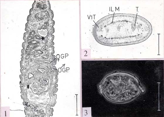
Lytocestus longicollis
| Fig. 1 : | Sagittal section through the posterior region revealing seperate male and female genital pores (scale bar= 0.15 mm) |
| Fig. 2 : | Transverse section showing the distribution of testes and vitellaria in relation to the longitudinal muscles (scale bar= 0.5 mm) |
| Fig. 3 : | Egg as seen under phase contrast (scale bar= 0.05) |